

Open any anatomy lab cupboard and the femur you lift out feels like pale wood: dry and light. Most of us carry that picture into the exam hall, and it's why the classification of bones, their composition and even their functions get filed under "lists to memorise". The picture is wrong in one way that matters. The femur in your thigh right now is wet, pink, fed by its own artery, and busy rebuilding itself.
Bone is a living organ, and that changes the whole topic
I wrote this for the student sitting with a disarticulated skeleton and a page of terms that all sound alike, because for my first month of osteology that student was me. I could recite bone names and still couldn't say what a bone actually was.
Bones are living, mineralised connective-tissue organs that make up the skeleton. An adult has 206 of them, and together they support the body, protect soft organs, act as levers for muscle, store calcium and phosphate, house the marrow that makes blood cells, and store fat. Their shape and surface markings reflect those jobs.
About that 206. Newborns carry roughly 270 bones, fusion trims the number, and StatPearls reports 206 to 213 in adults, the spread coming from anatomical variations such as extra small bones. Exams want 206: 80 in the axial skeleton (the long axis of the body, from skull down through the spine, plus the rib cage) and 126 in the appendicular skeleton (limbs and the girdles that attach them).
Here are the six jobs side by side.
| Function | What bone does | Where you meet it |
|---|---|---|
| Support | Rigid frame that carries body weight | Femur, vertebral column |
| Protection | Encloses soft organs | Skull around the brain, ribs around heart and lungs |
| Movement | Acts as a lever while muscle pulls on it | Humerus at the elbow, tibia at the knee |
| Mineral reservoir | Stores calcium and phosphate, releases them into blood on demand | Whole skeleton |
| Blood cell formation | Red marrow makes blood cells | Sternum, hip bone, upper ends of femur and humerus in adults |
| Fat storage | Yellow marrow holds an energy reserve | Medullary cavity of long bones |
Movement deserves a second look, because it explains half of what comes later. Bone can't move itself. Muscle pulls, and the bone turns around a joint like a lever around a pivot, which is why bones carry rough patches and ridges exactly where tendons attach. About 99% of the body's calcium sits in the skeleton, so bone is also the bank the blood draws on when levels dip. Newer research treats bone as an endocrine organ too, releasing signalling molecules, but most syllabi don't examine that yet, so file it as a bonus line.
Every one of those jobs depends on what bone is physically made of.
What is bone made of, and how is it built?
Bone is a composite: living cells embedded in a matrix that is roughly one-third organic (mostly type I collagen) and two-thirds mineral (hydroxyapatite, a calcium phosphate crystal, written Ca10(PO4)6(OH)2) by dry weight, with a little water. Proportions shift with age and site, so textbooks quote them as approximations.
Reinforced concrete is the closest everyday equivalent. Collagen plays the steel rods, because it stretches without snapping. Hydroxyapatite plays the concrete, because it resists crushing. Collagen buys toughness, mineral buys stiffness, and bone needs both.
The classic lab demonstration makes this concrete. Soak a bone in acid and the mineral dissolves; what's left bends like rubber. Heat one hard and the collagen burns away; the bone keeps its shape but crumbles at a touch.
Strip out one ingredient in real life and the bone tells you which one it was. Poor mineralisation gives soft bones that bend, the picture behind rickets in children and osteomalacia in adults (the causes include vitamin D and phosphate problems, which you'll meet in physiology and medicine). A faulty type I collagen gives bones that fracture easily, the story of osteogenesis imperfecta. Two conditions, opposite problems, one composite.
Four cell types run the matrix:
| Cell | Job |
|---|---|
| Osteoprogenitor cell | Stem-like cell that becomes an osteoblast |
| Osteoblast | Builds new matrix; once it's walled in by its own matrix it matures into an osteocyte |
| Osteocyte | Sits in a tiny pocket (lacuna) and senses mechanical load, keeping the matrix healthy |
| Osteoclast | Large cell that dissolves matrix (resorption), freeing calcium |
Builders and demolishers work side by side, which is why bone is never finished. StatPearls documents the practical result: bone resorbs when stress falls, so bedridden patients and astronauts on long flights lose bone over time. Load it and it thickens. Unload it and it thins.
Compact versus spongy
Here's the doubt nearly everyone has: if bone is "hard", why do pictures show holes inside it? Because bone comes in two textures. Compact bone is dense and solid, built from cylindrical units called osteons (concentric rings of matrix around a central canal that carries vessels and nerves). Spongy bone, also called cancellous bone, is a lattice of thin struts (trabeculae) with marrow in the gaps. The OpenStax account pairs each texture with a job: compact bone withstands compressive forces, while spongy bone copes with shifting weight distribution. A flat skull bone shows both at once, a spongy layer (the diploë) sandwiched between two compact plates.
Look at the struts inside the upper end of a femur and you'll see them lining up along the lines of force that bone normally carries, like girders placed by an engineer who knew where the load would go. That's remodelling doing the design work.
Inside a long bone
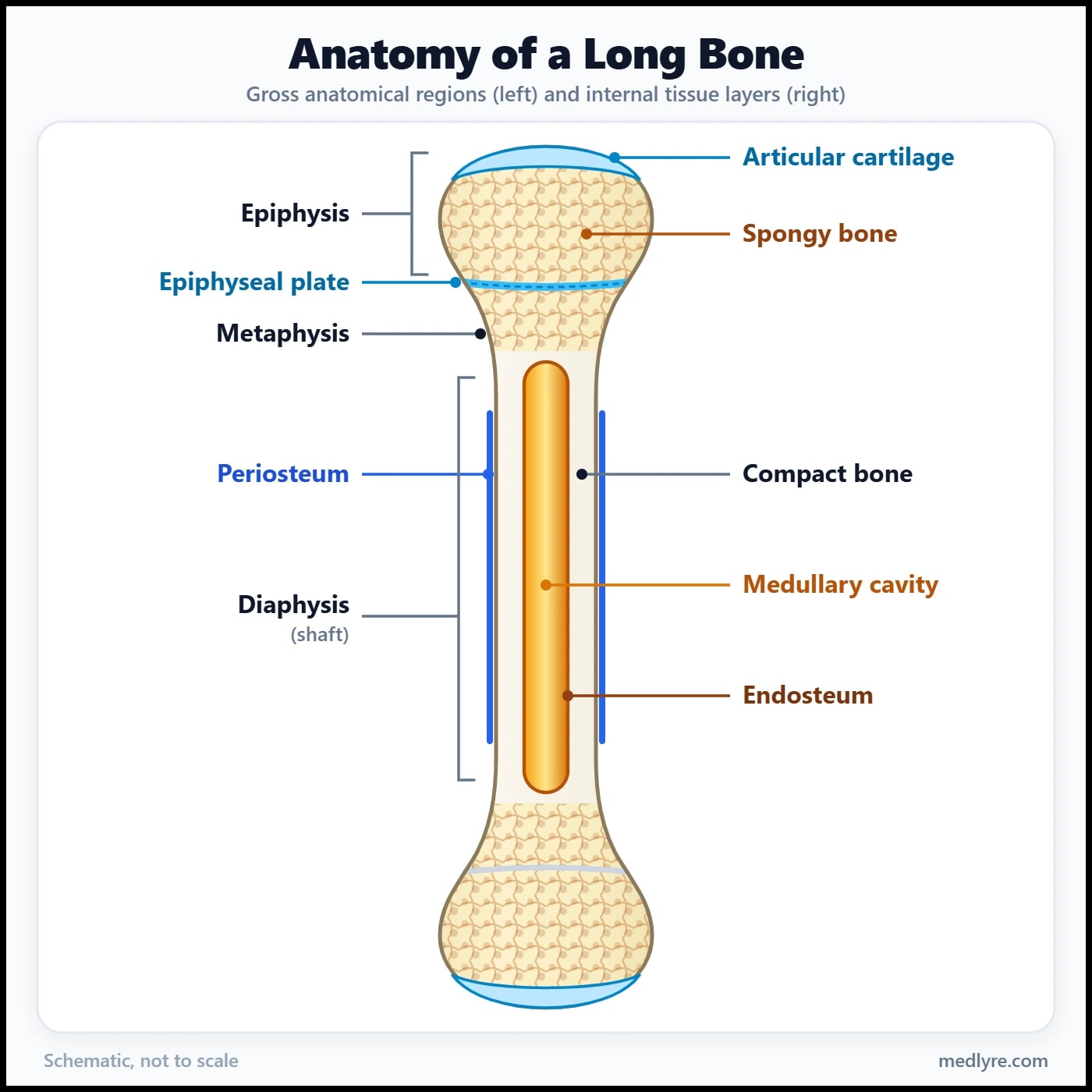
A long bone is the best model for labelling, since examiners love it.
| Part | What to remember |
|---|---|
| Diaphysis | The shaft. A thick tube of compact bone around the medullary cavity, which holds marrow. |
| Epiphysis | The expanded end. Spongy bone inside, a thin shell of compact bone outside, and articular cartilage where it meets another bone. |
| Metaphysis | The flared zone between shaft and end. In a growing child it lies right beside the growth plate. |
| Epiphyseal plate | A disc of hyaline cartilage that lengthens the bone through childhood and adolescence, then ossifies into the epiphyseal line. |
| Periosteum | Tough outer membrane with a fibrous layer and a bone-forming inner layer. Tendons anchor into it through Sharpey's fibres. |
| Endosteum | Thin lining of the medullary cavity and of the spongy bone's inner surfaces. |
Where students slip
Two traps live in this diagram. First, epiphysis and metaphysis get swapped constantly: the epiphysis is the end, the metaphysis is the flare behind it. Second, periosteum does not cover the joint surface. Articular cartilage does that job, which is why a bone end looks smooth and glassy where it meets its neighbour.
Next question: how do you sort 206 of these things?
Sorting 206 bones: shape first, then everything else
Shape is the classification of bones you'll be asked about most: long, short, flat, irregular and sesamoid, with sutural and pneumatic bones added by many university textbooks.
A long bone has a shaft and two ends and is longer than it is wide. Size is irrelevant. The femur qualifies, and so does a finger phalanx, which settles one of the most common doubts students have. Short bones are roughly cube-shaped, mostly spongy inside under a thin compact shell: the carpals and tarsals. Flat bones are thin and often curved, built as the sandwich you met earlier (the skull vault, sternum, ribs and scapula). Irregular bones fit no tidy shape, and the vertebrae are the standard example. Texts disagree on the hip bone, calling it flat or irregular, so irregular is the safer exam answer.
Sesamoid bones develop inside a tendon. The patella is the largest, the pisiform in the wrist sits in the tendon of flexor carpi ulnaris, and their shared trick is protecting the tendon from friction while changing its angle of pull. Pneumatic bones contain air-filled spaces, the sinuses of the frontal bone and maxilla being the usual examples. Sutural bones (also called Wormian bones) are small, variable extras that appear within skull sutures.
flowchart TD
    A["Bones classified by shape"] --> B["Long: shaft plus two ends (femur, phalanx)"]
    A --> C["Short: cube-like (carpals, tarsals)"]
    A --> D["Flat: compact plates around spongy bone (skull vault, sternum)"]
    A --> E["Irregular: no simple shape (vertebra)"]
    A --> F["Sesamoid: develops in a tendon (patella)"]
    A --> G["Pneumatic: air-filled spaces (maxilla)"]
    A --> H["Sutural: small, within skull sutures"]
Figure 2: The shape classification of bones, with one anchor example for each class.
One classic wrinkle: many textbooks describe the clavicle as a long bone that lacks a true medullary cavity, so expect that line in theory papers.
Shape isn't the only way to sort bones. Examiners reach for these schemes as well:
| Scheme | Groups | Example |
|---|---|---|
| By region | Axial (80) and appendicular (126) | Skull is axial; humerus is appendicular |
| By development | Membranous, cartilaginous, membrano-cartilaginous | Flat skull-vault bones form in membrane; limb long bones form from cartilage; the occipital bone has both |
| By internal structure | Compact and spongy | Shaft wall versus epiphysis |
The developmental split follows the same chapter's account of flat bones, which form by intramembranous ossification straight from mesenchyme, while long bones form by endochondral ossification on a cartilage model.
Exam pointer
When a question says "classify bones" with no qualifier, answer by shape and give one named example per class. Add region and development only if marks justify it.
Shape tells you what a bone looks like. The bumps and holes on its surface tell you what it does.
Reading the surface of a bone
A viva examiner once handed me a skull and said, "Don't just name the hole. Tell me who uses it." It changed how I read every bone afterwards.
The Oregon State adaptation of OpenStax groups bone markings into three classes: articulations, projections and holes. Every bump on a bone is a place something pulls, and every hole is a place something passes. Hold that line and half the terminology organises itself.
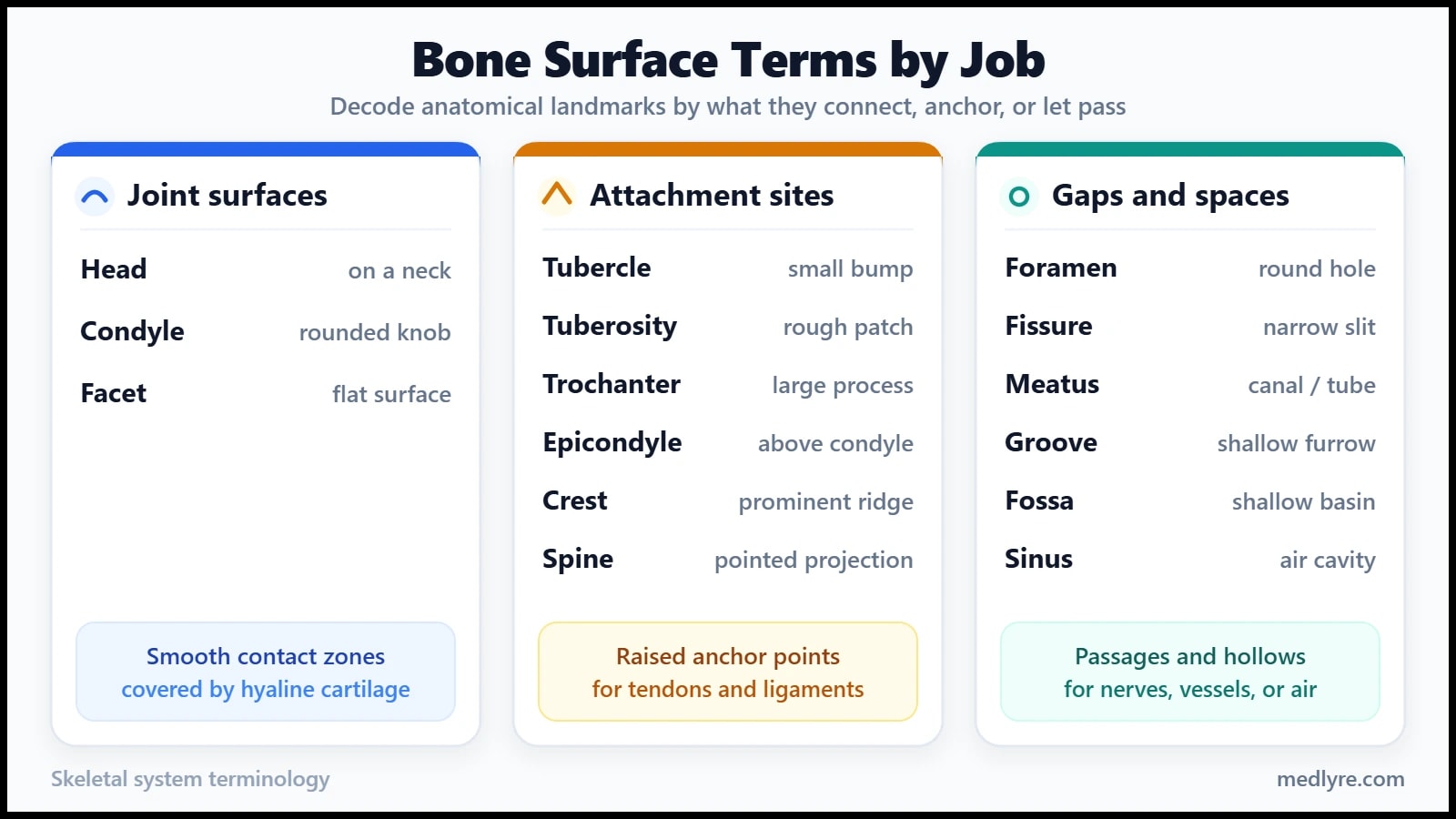
Projections are the group that confuses people, so take them as a ladder of size and roughness. A tubercle is a small rounded bump (greater tubercle of the humerus). A tuberosity is bigger and rougher (tibial tuberosity). A trochanter is the large blunt projection you meet on the femur. The boundaries are convention, not a measured cut-off, so don't argue with an examiner who calls something a tuberosity that your book calls a tubercle.
The condyle versus epicondyle pair works on the Greek prefix: epi means "upon", so the epicondyle sits on top of the condyle, doesn't form part of the joint, and serves as a muscle and ligament anchor. A head is a rounded articular end carried on a narrower neck, while a condyle is a knob without that neck. A crest is a prominent ridge (iliac crest), a line is a fainter one, and a spine is a sharp slender point. Keep "spine" (a pointed projection, as in the ischial spine) separate from the spinous process, which is the projection on the back of a vertebra.
The hole group has one rule: the passenger names the opening. A foramen is a round or oval hole, a fissure a narrow slit, a meatus a tube-like passage and a sinus an air-filled cavity. Kenhub offers a good worked case: the jugular foramen carries the glossopharyngeal, vagus and accessory nerves along with the inferior petrosal and sigmoid sinuses. A fossa is a shallow basin and a fovea a small pit (the fovea capitis of the femoral head). A groove or sulcus holds a tendon, vessel or nerve, and a notch (incisure) is an indentation in a bone's edge.
What breaks if a foramen narrows? Whatever passes through gets squeezed, which is why a narrowed intervertebral foramen can pinch the spinal nerve leaving it. That's the clinical reason these tiny words matter.
| Term | Plain meaning | Anchor example |
|---|---|---|
| Head | Rounded articular end on a neck | Head of femur |
| Condyle | Rounded articular knob | Femoral condyle, occipital condyle |
| Facet | Small, flat, smooth articular surface | Vertebral facet |
| Epicondyle | Non-articular bump above a condyle | Medial epicondyle of humerus |
| Tubercle | Small rounded projection | Greater tubercle of humerus |
| Tuberosity | Larger, rough projection | Tibial tuberosity |
| Trochanter | Large blunt projection | Greater trochanter of femur |
| Crest | Prominent ridge | Iliac crest |
| Spine | Sharp slender projection | Ischial spine |
| Foramen | Hole through bone | Jugular foramen |
| Fissure | Narrow slit | Superior orbital fissure |
| Meatus | Tube-like passage | External acoustic meatus |
| Groove | Furrow for a tendon, vessel or nerve | Bicipital (intertubercular) groove |
| Fossa | Shallow basin | Olecranon fossa |
Why it shows up in clinic
Radiology reports, fracture descriptions and surgical notes all locate things by these landmarks ("fracture through the greater trochanter", "compression at the foramen"). Fluent surface vocabulary means you can read a report instead of decoding it.
Ninety seconds before the viva
Pocket version
- Bone is a living organ: cells plus a matrix of about one-third organic material (mostly type I collagen) and two-thirds mineral (hydroxyapatite).
- Six jobs: support, protection, movement, mineral storage, blood cell formation, fat storage.
- Compact bone is dense and sits outside; spongy bone is a strut lattice inside; marrow fills the gaps and the medullary cavity.
- Long bone parts: epiphysis (end), metaphysis (flare beside the growth plate in children), diaphysis (shaft); periosteum outside, endosteum inside.
- Classification of bones by shape: long, short, flat, irregular and sesamoid, plus sutural and pneumatic.
- Surface terms sort into joint surfaces, attachment sites, and gaps and spaces.
- Foramen is a hole, fissure a slit, meatus a tube, fossa a basin, sinus an air-filled cavity.
Where students still get stuck
Q. How many bones are there in the adult human body?
The figure textbooks teach is 206, made up of 80 axial and 126 appendicular bones. Infants have around 270 because many bones are still separate pieces that fuse during growth. Real adults can differ slightly, since extra sutural bones or accessory bones vary from person to person, so a published range of 206 to 213 isn't a contradiction.
Q. Is a rib a long bone or a flat bone?
Flat. A rib is thin and curved, with spongy bone between two compact layers, and a long bone is defined by a shaft and two expanded ends. Ribs do look long in the everyday sense, which is exactly why this question keeps appearing in MCQs.
Q. What is the difference between a tubercle and a tuberosity?
Both are projections where tendons or ligaments attach. A tubercle is a small rounded bump, while a tuberosity is larger and has a rougher surface. The line between them is a matter of convention rather than a measured size, so learn the standard examples instead of arguing over cut-offs: the greater tubercle of the humerus and the tibial tuberosity. A trochanter, found on the femur, is the larger blunt version.
Q. Foramen, fissure, canal and meatus: how do I tell them apart?
Think of shape. A foramen is a round or oval hole, a fissure is a narrow slit, and a canal or meatus is a longer tube-like passage. Always name the passenger as well. If the examiner asks about the jugular foramen, the answer includes cranial nerves IX, X and XI.
Q. Why is the patella called a sesamoid bone?
Because it develops inside a tendon, here the quadriceps tendon, rather than as part of a bone shaft. It protects the tendon from friction over the knee and improves the angle at which the muscle pulls. The patella is the largest sesamoid, while tiny ones such as the pisiform sit in the wrist.
Q. If bone is so hard, why does it need blood vessels and cells?
Because it's alive and constantly remodelled. Osteoblasts build, osteoclasts remove, and osteocytes sense load, so the skeleton adapts to how you use it. Without blood supply, cells could not feed or repair the matrix, and the bone would behave like the dry specimen in the lab cupboard.
A note on scope
MedLyre articles are written for students learning the topic, not for patients making health decisions. Use this as a study aid alongside your textbooks and lectures. Clinical decisions belong in clinical settings, under qualified supervision, with full patient context.
References
- StatPearls Publishing. "Anatomy, Bones." StatPearls [Internet]. Treasure Island (FL): StatPearls Publishing; 2024 Jan-. PMID 30725884. https://www.ncbi.nlm.nih.gov/books/NBK537199/
- Betts JG, Young KA, Wise JA, et al. Anatomy and Physiology 2e. OpenStax; 2022. Section 6.3, "Bone Structure." https://openstax.org/books/anatomy-and-physiology-2e/pages/6-3-bone-structure
- Biga LM, Bronson S, Dawson S, et al. Anatomy & Physiology. Oregon State University / OpenStax; 2019. Section 7.2, "Bone Markings." https://open.oregonstate.education/anatomy/chapter/7-2-bone-markings/
- Kenhub. "Complete list of bone markings." Kenhub. https://kenhub.com/en/library/anatomy/complete-list-of-bone-markings