

Somewhere between 2 and 4 litres of fluid, depending on the source, leaks out of your capillaries every day and never makes it back through the veins. It takes plasma protein with it, and Guyton and Hall stress that this protein has no other route back. Without lymphatic return, plasma protein would drain into the tissues, plasma volume would keep falling until the circulation failed in shock, and death would follow within about a day, by Guyton's estimate. Something is doing that job right now, quietly, with no heart behind it.
Two to four litres a day: why the lymphatic system exists
Start at a single capillary. Near the arterial end, hydrostatic pressure shoves fluid out through the wall into the gap between cells (the interstitial space). Near the venous end, hydrostatic pressure has dropped enough for the oncotic pull of plasma proteins to win, and most of the fluid returns. Most. A small surplus stays behind, and so do a few protein molecules that slipped through the wall and are too large to be reabsorbed.
The first time I tried to explain this to a junior, I realised I had memorised the node groups without ever asking where one drop of fluid actually went. So this article follows a single drop from the tissue to the vein, and the aim is simple: by the end you can redraw the whole route from memory on a blank page.
The lymphatic system is a one-way drainage network. It collects excess tissue fluid and leaked protein, filters that fluid through lymph nodes, and returns it to the blood at the venous angles. It also houses immune cells, so it works as a recycling line and a surveillance system at once.
Why does the protein matter so much? Guyton and Hall put the amount leaking out of blood capillaries at roughly 100 grams a day. Water and small solutes follow pressure gradients back into venous capillaries. Large protein molecules can't. The lymphatic system is the return line for everything the veins cannot take back.
Follow what happens without it. Protein piles up in the tissue, so the oncotic pull of the interstitial fluid rises, so more water gets drawn out of the blood, so the tissue swells further. Each step feeds the next. That runaway swelling is oedema, and it is what a failed return looks like. First, though, the fluid has to get inside a vessel that has no open end.
One-way flaps at the start of the line
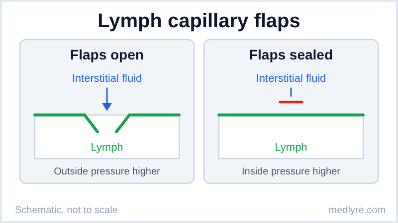
A lymph capillary begins as a blind-ended tube. Its wall is a single layer of endothelial cells whose edges overlap like roof tiles, and fine anchoring filaments tether those cells to the surrounding tissue. When fluid builds up, the tissue swells, the filaments pull, and the overlapping edges swing inward. Fluid pours in. The moment pressure inside the vessel rises above the pressure outside, the edges press shut, so nothing leaks back out.
Imagine a letterbox flap on a front door. Post goes in easily, and nothing comes back out, because the flap only swings one way. Figure 1 shows the same trick at capillary scale.
Moore's Clinically Oriented Anatomy describes lymph capillaries as beginning blindly in the tissue spaces, and notes that they are absent from avascular tissues such as the epidermis and cartilage. Older texts add the brain and spinal cord to that list. A 2015 Nature paper by Louveau and colleagues described lymphatic vessels in the meninges, so treat "no lymphatics in the CNS" as classic teaching that is under revision.
Where students slip
Many students sketch lymph capillaries as tiny veins that join arteries. They never join arteries. They start blind in the tissue spaces, which means fluid can only enter, and the first real connection to the blood is the big duct at the far end of the route.
The gut adds a twist. Each intestinal villus holds a central lymph capillary called a lacteal. Absorbed fat, packed into chylomicrons, is too bulky to enter blood capillaries, so it slides into the lacteal instead and turns gut lymph milky. That milky lymph is chyle. Standard physiology texts trace absorbed fat into the lacteals this way.
Fluid is now inside the vessel. The next puzzle is propulsion.
How does lymph climb back up without a heart?
Blood has a heart. Lymph has nothing of the kind, yet it travels from your ankle to the base of your neck. The answer is a handful of small forces working together, and they split into two groups.
The first group sits inside the vessel. Collecting lymph vessels are divided into segments by valves, and each segment (a lymphangion) has smooth muscle in its wall. When a segment stretches with incoming lymph, the muscle contracts and pushes the lymph forward. The valve behind it snaps shut. Repeat that down the chain and you have a line of tiny pumps. Guyton and Hall detail this valved, self-contracting arrangement as the lymphatic system's own pump.
The second group sits outside. Skeletal muscle squeezes vessels as it contracts, arteries pulse beside them, and breathing joins in: each inspiration lowers pressure in the chest and helps draw lymph toward the thoracic duct. Because the valves allow only one direction, every squeeze from outside acts as a push forward.
| Force | Where it acts | Concrete example |
|---|---|---|
| Lymphangion smooth muscle | Inside collecting vessels | Stretch from incoming lymph triggers contraction |
| Skeletal muscle contraction | Outside the vessel | Calf muscles compressing deep lymphatics with each step |
| Arterial pulsation | Adjacent vessels | A deep lymph vessel running beside an artery |
| Respiratory movement | Thoracic region | Inspiration lowering chest pressure around the thoracic duct |
Quick check before moving on: what stops lymph from sliding backwards between pushes? The valves. The forces generate movement, and the valves turn that movement into one-way flow.
What controls how much lymph moves? Interstitial fluid volume does. Guyton and Hall's flow graph shows lymph flow rising as interstitial pressure rises, then levelling off, because a very swollen tissue starts squeezing its own lymph vessels. The same source puts total lymph flow near 120 mL an hour, with about 100 mL an hour of that passing through the thoracic duct. Multiply that out over 24 hours and you get about 2.9 litres, inside the 2 to 4 litre range from the start of this article.
Fluid that has travelled this far has still not met a single filter. That comes next.
Nodes filter, ducts deliver: the last stretch to the neck
A lymph node is a bean-shaped filter, from about a millimetre to a couple of centimetres long. Several afferent vessels pierce its convex surface, and one or two efferent vessels leave at the hilum. Afferent arrives, efferent exits. Many in and few out means lymph slows down inside the node, and slow flow buys time for inspection.
Inside, lymph enters the subcapsular sinus, passes through the trabecular (cortical) sinuses, and reaches the medullary sinuses before leaving by the efferent vessel. Macrophages line the subcapsular and medullary sinuses and engulf debris and microbes as lymph flows past. Antigen and cells move from the sinuses into the surrounding lymphoid tissue: B-cell follicles sit in the outer cortex, while T cells and dendritic cells gather in the paracortex, where the dendritic cells present antigen to T lymphocytes. Plasma cells fill the medullary cords. Robbins and Cotran lays out this compartment map as the architecture behind reactive node enlargement.
Nodes belong to a wider family. The thymus and bone marrow are the primary lymphoid organs, where lymphocytes are made and trained. Nodes, the spleen, tonsils, and the lymphoid patches of the gut are secondary organs, the sites where immune cells encounter antigen. The spleen does for blood roughly what nodes do for lymph.
After the nodes, efferent vessels merge into larger lymphatic trunks, and the trunks feed two ducts. Figure 2 shows the whole route as a single chain.
flowchart TD
    A["Interstitial fluid"] -->|"pressure opens flaps"| B["Lymph capillary"]
    B -->|"valves keep flow forward"| C["Collecting vessel"]
    C -->|"afferent vessels"| D["Lymph node"]
    D -->|"efferent vessel"| E["Lymphatic trunk"]
    E -->|"merges into"| F["Thoracic duct or right lymphatic duct"]
    F -->|"drains into"| G["Venous angle"]
Figure 2: The route one drop of lymph takes from tissue to vein.
The thoracic duct starts in the upper abdomen as a sac called the cisterna chyli (around L1 to L2, according to Moore), climbs through the aortic opening of the diaphragm, and empties at the left venous angle, where the left subclavian and left internal jugular veins meet. The right lymphatic duct is short and serves a smaller territory.
| Duct | What it drains | Where it empties |
|---|---|---|
| Thoracic duct | Everything below the diaphragm, plus the left arm, the left side of the head and neck, and the left chest | Left venous angle |
| Right lymphatic duct | Right arm, right side of the head and neck, right chest | Right venous angle |
Contrast is the anchor here: the thoracic duct carries the heavy load, and the right duct handles one upper quadrant only. A student who remembers that everything below the diaphragm drains to the left venous angle has the core of the answer.
The nodes you can actually feel on a body map onto this plan by region. Moore's Clinically Oriented Anatomy groups them so that the cervical nodes serve the head and neck, the axillary nodes serve the upper limb, the breast, and the upper trunk wall, and the superficial inguinal nodes serve the lower limb, the perineum, and the lower abdominal wall. Each group acts as the first filter for its own territory, and the axilla will matter again in the next section.
That completes the route. Now break it and see what happens.
What breaks when lymph drainage is blocked?
What happens if one stretch of the route is blocked? Capillaries still filter fluid on schedule, because filtration depends on blood pressures, not lymph. Nothing carries the surplus away, so it accumulates. The protein trapped with it raises the interstitial oncotic pull, which draws in still more water. This is lymphoedema: protein-rich swelling that, over months, turns firm as fibrosis (scar-like tissue) sets in.
Robbins and Cotran lists the classic causes of obstruction: tumour, surgery, radiation, and infection of the vessels themselves. Surgery gives the cleanest teaching example. Removing axillary nodes during breast cancer surgery cuts the arm's main drainage route, so arm lymphoedema is a recognised consequence.
Parasites give the tropical example. Adult Wuchereria bancrofti worms lodge in lymphatic vessels, provoke inflammation, and over years scar the channels shut. The outcome is massive limb or scrotal enlargement, the picture called elephantiasis, and it's a staple of pathology and community-medicine teaching.
Why this shows up in clinic
Nodes themselves can enlarge. When an infection floods a region, lymphocytes multiply inside the draining nodes and the nodes swell. Textbooks teach students to read an enlarged node by its location and by how it feels (tender or painless, mobile or fixed), which helps separate a reactive node from other causes.
That covers the whole system. The card below compresses it.
Lymph drainage on one index card
Revision snapshot
- The lymphatic system returns about 2 to 4 litres of fluid a day, plus protein that blood capillaries can't take back.
- Lymph capillaries are blind-ended, with overlapping endothelial flaps held open by anchoring filaments.
- Lymph moves through valved vessels powered by lymphangion contraction, skeletal muscle, arterial pulsation, and breathing.
- Afferent vessels enter a node on the convex side (many); the efferent vessel leaves at the hilum (one or two).
- Thoracic duct: everything below the diaphragm plus the left upper quadrant, into the left venous angle. Right lymphatic duct: right upper quadrant, into the right venous angle.
- Blocked drainage means protein-rich swelling (lymphoedema); gut lymph is milky chyle carrying chylomicrons.
Loose ends students raise after the lymph lecture
Q. What is the difference between lymph and plasma?
Lymph starts life as interstitial fluid, so it resembles plasma but usually carries far less protein, because only a little protein leaks through blood capillary walls. It also contains lymphocytes, and after a fatty meal, gut lymph carries chylomicrons. Plasma stays inside blood vessels. Lymph forms outside them, travels in its own vessels, and rejoins the blood at the venous angles.
Q. Does the lymphatic system have its own pump?
Not a single central pump like the heart, but it has local ones. Collecting vessels are divided into lymphangions, segments with smooth muscle and valves, and each segment contracts when stretched. Skeletal muscle activity, arterial pulsation, and breathing add outside pressure. The valves make sure all that pushing moves lymph in one direction only, toward the neck.
Q. Why does the thoracic duct drain more of the body than the right lymphatic duct?
It collects from everything below the diaphragm and from the left side above it. The right lymphatic duct serves only the right upper quadrant: the right arm, the right side of the head and neck, and the right chest. Add the entire lower body to the left upper quadrant, and the thoracic duct's territory wins by a wide margin.
Q. What is chyle?
Chyle is the milky lymph that drains the small intestine after a fatty meal. Absorbed fat leaves the intestinal cells packaged as chylomicrons, which are too large to enter blood capillaries, so they enter the central lacteal of each villus. The lacteals feed collecting vessels that gather at the cisterna chyli, where the thoracic duct begins its climb.
Q. What is lymphoedema in simple words?
Lymphoedema is swelling caused by fluid and protein that can't leave the tissue because lymph drainage is blocked or damaged. Capillaries keep filtering fluid, but the return route is closed. Protein builds up and pulls more water in, so the swelling grows. Over time, the tissue stiffens as fibrosis develops. Node surgery and tumour obstruction are the usual teaching examples, and filariasis is the classic parasitic cause.
Q. Why do lymph nodes swell during an infection?
Antigens drain from the infected area to the nearest nodes, where lymphocytes meet them, activate, and multiply. B-cell follicles expand and the node fills with proliferating cells, so it enlarges. That is called reactive enlargement. Because lymph drains region by region, the location of the swollen node points back toward the area that sent the antigen.
For students, not patients
Everything in this article exists to help students understand a topic they struggle with. It is not a treatment guide, not a clinical protocol, and not a substitute for the curriculum your institution follows. Bedside decisions always need qualified supervision and full patient assessment.
References
- Hall JE, Hall ME. Guyton and Hall Textbook of Medical Physiology. 14th ed. Elsevier; 2020.
- Barrett KE, Barman SM, Brooks HL, Yuan JXJ. Ganong's Review of Medical Physiology. 26th ed. McGraw-Hill; 2019.
- Moore KL, Dalley AF, Agur AMR. Clinically Oriented Anatomy. 8th ed. Wolters Kluwer; 2018.
- Kumar V, Abbas AK, Aster JC. Robbins and Cotran Pathologic Basis of Disease. 10th ed. Elsevier; 2020.
- Louveau A, Smirnov I, Keyes TJ, et al. "Structural and functional features of central nervous system lymphatic vessels." Nature. 2015;523(7560).