

Your heart is striped. Put a slice of it under a microscope and the banding looks almost identical to a slice of your biceps, line for line. Yet you can flex that biceps on command, and you can't tell the heart to skip a single beat. Same stripes, opposite control.
That mismatch is where most answers on voluntary and involuntary muscles go wrong, and it's the reason examiners keep coming back to this topic.
Voluntary and involuntary muscles sort by control alone
Voluntary muscles are the ones you can contract on purpose, and in the human body that means skeletal muscle. Involuntary muscles, cardiac and smooth muscle, contract without conscious command, driven by pacemaker cells, the autonomic nervous system, hormones and local signals. Voluntary and involuntary describe control, while striated and smooth describe appearance.
In my own first-year notes I filed the heart under "voluntary" for weeks, purely because it looked so much like skeletal muscle on the slide. A viva question finally exposed the mistake. I wrote this article to pull that knot apart for you: two separate questions get squeezed into a single label, and once you ask them one at a time, the three muscle types click into place.
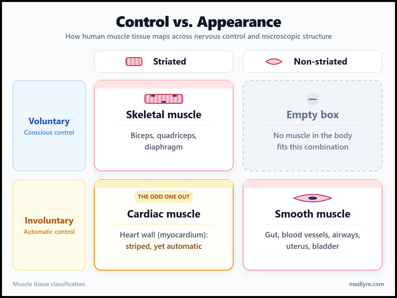
Question 1 is about control. Can you decide to contract the muscle? Voluntary means yes, involuntary means no, and the answer comes from the wiring of the nervous system and from the cell's own rhythm. Question 2 is about appearance. Does the fibre show cross-stripes under a microscope? Striated means yes, smooth means no, and the stripes come from actin and myosin being lined up into repeating units called sarcomeres.
Cross the two answers and you get a small grid. Skeletal muscle is striated and voluntary. Cardiac and smooth muscle are both involuntary, but only cardiac wears the stripes. The fourth box, non-striated and voluntary, stays empty in the human body.
Where students slip
Writing "striated muscle is voluntary" costs marks, because cardiac muscle is striated and involuntary. Striated describes the look of the fibre. Voluntary describes who is in control. Keep them in separate columns in your head and the heart stops being a puzzle.
Honestly, if I had to pick one idea from this whole topic to carry into the exam hall, it would be this two-axis grid. So what does the one muscle you can actually command look like on the inside?
Skeletal muscle: from the whole biceps down to one sarcomere
Skeletal muscle is the voluntary, striated muscle that pulls on your bones, and it's the heavyweight of the body. The StatPearls physiology review puts it at about 40% of body weight and 50 to 75% of all body protein.
Hold a rope in your mind and you already own the architecture. Thin strands twist into thicker strands, thicker strands into the rope, and every level has its own wrapping. A muscle is built the same way, from the outside in. The NCBI Bookshelf chapter on skeletal muscle anatomy lays out the outermost sheath around the whole muscle as the epimysium, with the bundles inside it called fasciculi.
| Level | Boundary or wrapper | What it holds |
|---|---|---|
| Whole muscle (the biceps) | Epimysium | Many fascicles |
| Fascicle | Perimysium | A bundle of muscle fibres |
| Muscle fibre (one cell) | Sarcolemma, with endomysium outside it | Hundreds to thousands of myofibrils |
| Myofibril | Sleeve of sarcoplasmic reticulum | A chain of sarcomeres |
| Sarcomere | Z disc at each end | Thin actin and thick myosin filaments |
One fibre is one cell, and it's a giant. During development many myoblasts fuse into a single long cell, which is why a mature fibre is multinucleate and keeps its nuclei pushed to the edge, just under the sarcolemma. A stained skeletal slice shows flattened nuclei hugging the border of every fibre.
Inside the fibre, myofibrils run lengthwise like parallel cables, and each cable is a chain of sarcomeres joined end to end at the Z discs. Thin actin filaments hang from the Z discs while thick myosin filaments sit in the middle. The region holding the thick filaments forms the dark A band, the zone with only thin filaments forms the pale I band, and that repeating pattern is what you see as stripes. When the muscle contracts, thin filaments slide inward over the thick ones, the sarcomere shortens, and the I band narrows while the A band keeps its width.
How does a nerve signal reach all that machinery? The action potential spreads along the sarcolemma and dives into the fibre through the T-tubules, which run between 2 terminal cisternae of the sarcoplasmic reticulum at the junction of the A and I bands. That arrangement is called a triad. The sarcoplasmic reticulum releases calcium, calcium binds troponin C on the thin filament, tropomyosin shifts aside, and myosin finds its binding sites.
The word "voluntary" comes from the wiring. Each fibre sits under one neuromuscular junction from a somatic motor neuron, and a neuron plus every fibre it supplies makes a motor unit. Guyton and Hall point out that muscles needing fine control, such as some laryngeal muscles, run as few as 2 or 3 fibres per motor unit, while large postural muscles can run to several hundred. That ratio is why your eyes steer with precision and your gluteals shove with power.
When a voluntary muscle acts on its own
Here's the doubt that sneaks up on people. Voluntary describes what the muscle can do, and it never promises that every contraction is deliberate. Tap just below your kneecap and the quadriceps fires before you've decided anything. Fall asleep and the diaphragm keeps going, driven by the brainstem. Shivering recruits skeletal fibres purely to make heat. All 3 are skeletal muscle doing involuntary work, so the right test is whether conscious control is available through the somatic nervous system.
The Bookshelf's muscle physiology chapter lists striated muscle in places students forget: the upper oesophagus, the external anal sphincter and the external urethral sphincter. The smooth muscle of the bladder wall works without asking you, while the striated external sphincter waits for your decision.
Skeletal muscle also repairs itself reasonably well, because satellite cells lying against each fibre can divide and fuse into it after damage.
Everything so far assumed a muscle waits for a nerve to speak. The next muscle starts its own conversation.
Why can't the heart ever cramp?
Cardiac muscle can't go into sustained tetanus, because its long action potential keeps the cell refractory for the whole contraction. A ventricle locked mid-squeeze would be useless, so the design makes that physically impossible.
Under the microscope, cardiomyocytes are short, branched and striped, with usually 1 nucleus sitting in the middle of the cell. They carry the same sarcomere machinery as skeletal fibres, which explains the matching stripes. Their T-tubules sit at the Z discs, and OpenStax documents that this leaves cardiac muscle with about half as many T-tubules as skeletal muscle, which has them at both A-I junctions of every sarcomere.
The feature that gives cardiac muscle away is the intercalated disc, the stepped junction where one cell's end meets the next. Fascia adherens and desmosomes handle the mechanics: the first anchors the actin of the last sarcomere to the membrane so force passes from cell to cell, and the second rivets neighbours together against the pull of every beat. Gap junctions handle the electricity by opening small channels so ions cross directly from cytoplasm to cytoplasm.
Rivets and open doorways make a fair everyday picture here, the doorways being the gap junctions left open between neighbouring rooms. Current flows through those doorways, so an impulse that starts in one cell reaches its neighbours almost at once. Guyton and Hall describe the result as 2 functional syncytia, one atrial and one ventricular, each contracting as a single unit.
Dissolve the gap junctions and the ventricle becomes a crowd of cells, each beating to its own time. No coordinated squeeze, no pressure build-up, no ejection.
The heart also writes its own script. Pacemaker cells in the sinoatrial node generate impulses without waiting for a nerve, and the autonomic nervous system and hormones only speed or slow a rhythm that already exists. Calcium handling follows a similar pattern: calcium entering through the sarcolemma during the plateau triggers a larger release from the sarcoplasmic reticulum, and then the same troponin C switch used in skeletal muscle takes over.
Now the question in the heading. In skeletal muscle, a StatPearls chapter on skeletal muscle physiology explains, the action potential ends before the twitch does, so a second signal can arrive while the fibre is still contracting and the twitches add up into tetanus. Pull the long refractory period out of the cardiac design and the same summation would hit the ventricle. Medicine LibreTexts reminds us that tetany in the heart is incompatible with life, since the chambers could neither fill nor eject blood.
The numbers make the point. The same textbook gives a skeletal action potential roughly 1 to 5 ms, while a ventricular one lasts a couple of hundred. The refractory period outlasts the contraction, so there's no moment when a second contraction can pile onto the first. That one fact is the bit that still gets me every time I draw the two curves side by side.
Next: the muscle with no stripes at all.
Smooth muscle, built without sarcomeres
Smooth muscle is the involuntary, non-striated muscle of the gut, blood vessels, airways, uterus, bladder and iris, and it contracts with actin and myosin that aren't arranged into sarcomeres.
A classmate once asked me whether goosebumps were muscle or just skin reacting to cold. Muscle: tiny arrector pili bundles, which are smooth muscle, tug each hair upright.
Each smooth muscle cell is a spindle, wide in the middle and tapering at both ends, with a single central nucleus. Medicine LibreTexts sketches these cells at roughly 30 to 200 μm and likens the dense body, the dark anchor that holds the thin filaments, to the Z disc of striated muscle. Thin filaments run obliquely across the cell, tied to dense bodies on the membrane and to a mesh of intermediate filaments. A contraction therefore pulls both ends toward the middle and the cell bulges like a corkscrew. Without lined-up sarcomeres there are no stripes.
Smooth muscle has no troponin, so what does the calcium switch? OpenStax traces the job to calmodulin: calcium arriving from outside the cell and from the sarcoplasmic reticulum binds calmodulin, the calcium-calmodulin pair activates myosin light chain kinase (MLCK), and MLCK phosphorylates the myosin head so it can grab actin. Skeletal muscle works like a gate that swings open when calcium arrives. Smooth muscle works like a relay: calcium passes the message to calmodulin, calmodulin to an enzyme, and the enzyme tags myosin with a phosphate.
flowchart TD
    A["Ca²⁺ rises inside the cell"] -->|"influx and SR release"| B["Ca²⁺ binds calmodulin"]
    B -->|"activates enzyme"| C["Calmodulin activates MLCK"]
    C -->|"adds phosphate"| D["MLCK phosphorylates myosin light chain"]
    D -->|"head switches on"| E["Phosphorylated myosin binds actin"]
    E -->|"ATP splitting"| F["Cross-bridge cycling builds tension"]
    F -->|"stimulus ends"| G["Ca²⁺ falls, MLCP removes the phosphate"]
    G --> H["Relaxation"]
Figure 2: The smooth muscle calcium switch, from rising calcium to relaxation.
The pathway ends when calcium falls and myosin light chain phosphatase (MLCP) strips the phosphate off, a step the point-of-care smooth muscle entry outlines as the trigger for relaxation.
Why a blood vessel can hold tone cheaply
Lumen Learning's smooth muscle chapter attributes this to latch-bridges: a subset of cross-bridges stays linked for long periods with very little energy cost. That's how arterioles and hollow organs keep steady tone all day without tiring.
Control comes from the autonomic nervous system, hormones and local chemistry, and visceral smooth muscle also responds to stretch. Nerve endings reach the cells through varicosities, swellings along the axon that release transmitter onto several cells, and the same chapter confirms that no organised neuromuscular junction forms. That's why smooth muscle contracts slowly and for a long time, ideal for pushing food along or narrowing a vessel.
Gap junctions split smooth muscle into 2 working styles. Single-unit smooth muscle, found in the gut and most blood vessels, links its cells so a wave spreads and the sheet contracts as one. Multi-unit smooth muscle, found around large airways, in the iris and around large vessels, rarely has gap junctions, so each cell responds to its own input and a contraction stays where it started.
Unlike skeletal and cardiac muscle, smooth muscle can add cells by dividing. The uterus grows this way at puberty, when rising oestrogen drives new fibres into the myometrium.
Put them under the microscope
Slide questions reward a fixed routine of 2 steps. First ask: are there stripes? None means smooth muscle, a spindle cell with one central nucleus, and you're done. Stripes mean skeletal or cardiac, so ask second: where are the nuclei, and do the fibres branch? Many nuclei crowded along the edge of long parallel fibres mean skeletal. One central nucleus, branching fibres, and dark stepped lines crossing the fibre mean cardiac, because those lines are the intercalated discs.
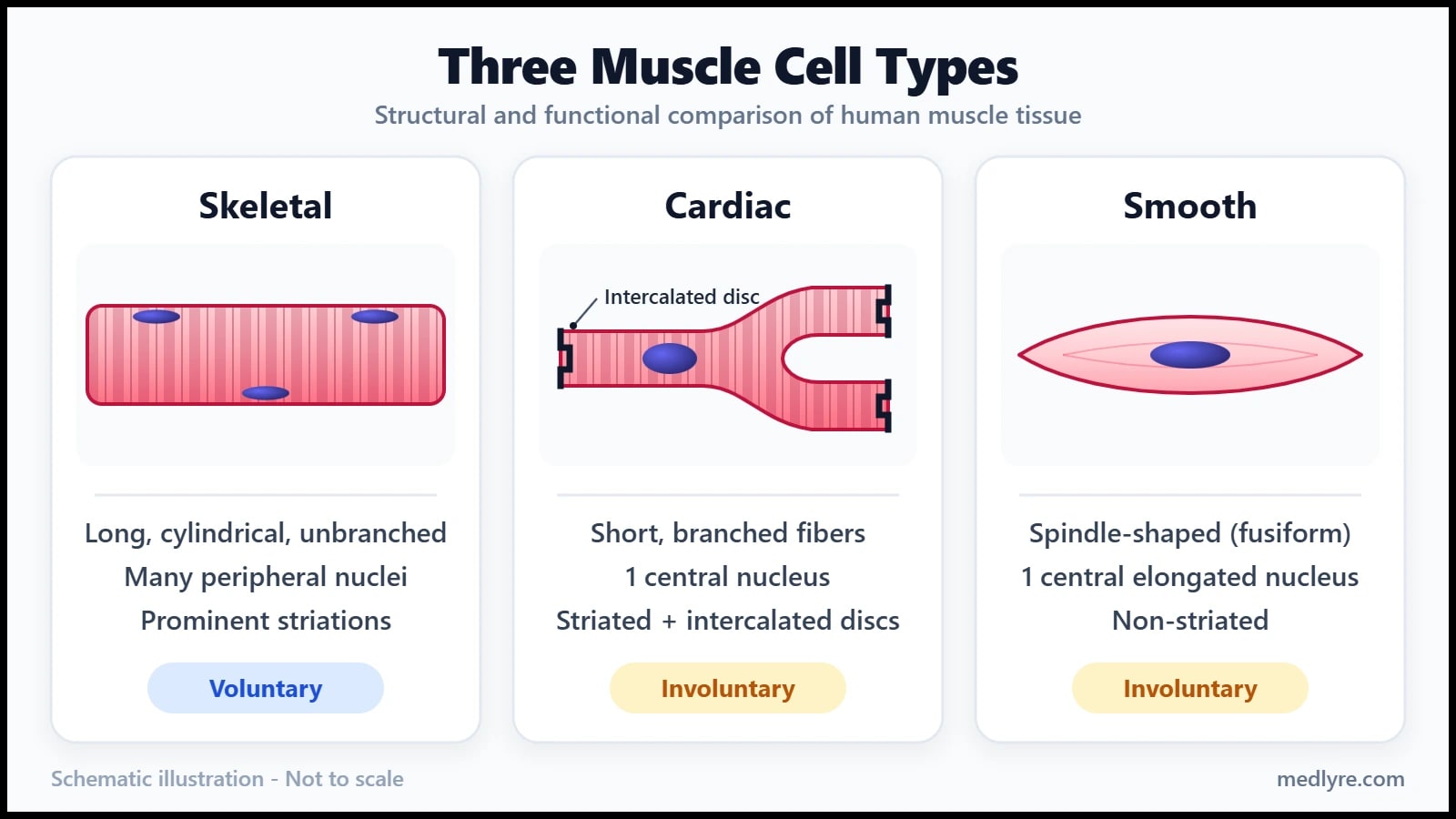
Keep this table beside you when you revise, because it holds almost every comparison an examiner can ask.
| Feature | Skeletal | Cardiac | Smooth |
|---|---|---|---|
| Control | Voluntary, via somatic motor neurons | Involuntary, own pacemaker, tuned by autonomic nerves and hormones | Involuntary, autonomic nerves, hormones, local factors, stretch |
| Cell shape | Long, unbranched cylinder | Short, branched | Spindle |
| Nuclei | Many, at the edge | Usually 1, central | 1, central |
| Stripes | Yes | Yes | None |
| Sarcomeres | Yes | Yes | No, dense bodies anchor the filaments |
| Junctions between cells | None, each fibre works alone | Intercalated discs (fascia adherens, desmosomes, gap junctions) | Gap junctions in the single-unit type |
| T-tubules | At A-I junctions | At Z discs | None, shallow caveolae instead |
| Calcium switch | Troponin C | Troponin C | Calmodulin and MLCK |
| Contraction pattern | Fast, can summate to tetanus | Rhythmic twitch, can't tetanise | Slow and sustained |
| Repair | Satellite cells | Very limited, scar replaces dead cells | Cells can divide |
Ninety seconds before the viva
Say these out loud before you walk in
- Voluntary and involuntary muscles are sorted by control. Striated and smooth are sorted by appearance. Keep them on separate axes.
- Skeletal: voluntary, striated, many edge nuclei, troponin C switch, force graded by motor-unit recruitment.
- Cardiac: involuntary, striated, 1 central nucleus, intercalated discs, own pacemaker, can't tetanise because the refractory period outlasts the contraction.
- Smooth: involuntary, no stripes, spindle cell, calmodulin and MLCK, slow and sustained, single-unit or multi-unit.
- Two traps: striated doesn't mean voluntary, and skeletal muscle can act involuntarily (knee jerk, sleeping diaphragm, shivering).
Where do students still slip after the first read?
Q. Is every skeletal muscle voluntary all the time?
Skeletal muscle is voluntary in the sense that conscious control is available through the somatic motor system, and a lot of its activity still runs without your decision. The knee jerk, the diaphragm during sleep, and shivering all use skeletal fibres automatically. Exam answers stay safe when they say skeletal muscle is under voluntary control but also acts reflexly.
Q. Why is cardiac muscle involuntary when it looks like skeletal muscle?
Appearance and control are separate axes. Cardiac cells share the sarcomere pattern that produces stripes, so they look like skeletal fibres, yet their rhythm starts in pacemaker cells of the sinoatrial node and spreads through gap junctions. No conscious command enters the loop, and the autonomic nervous system only speeds or slows the beat.
Q. Does involuntary mean a muscle has no nerve supply?
Involuntary muscles carry plenty of nerves. Smooth muscle receives autonomic fibres that end in varicosities, and the heart receives sympathetic and parasympathetic input that tunes a rate the pacemaker already sets. Hormones and local chemicals act on both. Involuntary describes the absence of conscious command, and the circuitry behind it stays rich.
Q. Which muscle types have sarcomeres?
Skeletal and cardiac muscle do, which is why both look striped. Smooth muscle has actin and myosin but no sarcomeres, so its thin filaments anchor to dense bodies, the counterparts of Z discs, and the cell shows no stripes. Sarcomeres give stripes, and calmodulin replaces troponin when they're absent.
Q. How do I tell cardiac from skeletal muscle on a slide?
Check the nuclei first. Skeletal fibres are long, unbranched and carry many nuclei pressed against the edge. Cardiac fibres branch, usually hold 1 nucleus in the middle, and show dark stepped intercalated discs running across the fibre. If you see branching, central nuclei and those steps, write cardiac.
Q. Which muscle repairs itself best?
Smooth muscle can add cells by dividing, and skeletal muscle repairs through satellite cells that fuse into damaged fibres. Cardiac muscle repairs poorly, which is why dead tissue after a heart attack gets replaced by scar rather than new contractile cells. That limited repair is a favourite one-line answer in short-note questions.
For students, not patients
Everything in this article exists to help students understand a topic they struggle with. It's not a treatment guide, not a clinical protocol, and not a substitute for the curriculum your institution follows. Bedside decisions always need qualified supervision and full patient assessment.
References
- Hall JE, Hall ME. Guyton and Hall Textbook of Medical Physiology. 14th ed. Elsevier; 2020.
- StatPearls Publishing. "Physiology, Skeletal Muscle." StatPearls [Internet]. Treasure Island (FL); 2026 Jan-. PMID: 30725824. Direct URL.
- StatPearls Publishing. Skeletal muscle physiology chapter. StatPearls [Internet]. NCBI Bookshelf ID NBK559006. Direct URL.
- StatPearls Publishing. Skeletal muscle anatomy chapter. StatPearls [Internet]. NCBI Bookshelf ID NBK537236. Direct URL.
- StatPearls Publishing. Muscle physiology chapter. StatPearls [Internet]. NCBI Bookshelf ID NBK537140. Direct URL.
- StatPearls Publishing. "Physiology, Smooth Muscle." Editor: Bracken Burns. Updated 2023. Direct URL.
- OpenStax. Anatomy and Physiology. "Cardiac Muscle and Electrical Activity." Lumen Learning edition. Direct URL.
- OpenStax. Anatomy and Physiology. "10.8 Smooth Muscle." Direct URL.
- Medicine LibreTexts. "17.3: Cardiac Muscle." Skyline College, BIOL 250 Human Anatomy. Direct URL.
- Medicine LibreTexts. "9.7: Smooth Muscle." Skyline College, BIOL 250 Human Anatomy. Direct URL.
- Lumen Learning. "Smooth Muscle." Anatomy and Physiology I (Trident Technical College). Direct URL.